| Fachbereiche | |
|---|---|
| Buchreihen (97) |
1382
|
| Nachhaltigkeit |
3
|
| Gesundheitswesen |
1
|
| Geisteswissenschaften |
2372
|
| Naturwissenschaften |
5408
|
| Mathematik | 228 |
| Informatik | 320 |
| Physik | 980 |
| Chemie | 1364 |
| Geowissenschaften | 131 |
| Humanmedizin | 243 |
| Zahn-, Mund- und Kieferheilkunde | 10 |
| Veterinärmedizin | 108 |
| Pharmazie | 147 |
| Biologie | 835 |
| Biochemie, Molekularbiologie, Gentechnologie | 121 |
| Biophysik | 25 |
| Ernährungs- und Haushaltswissenschaften | 45 |
| Land- und Agrarwissenschaften | 1005 |
| Forstwissenschaften | 201 |
| Gartenbauwissenschaft | 20 |
| Umweltforschung, Ökologie und Landespflege | 148 |
| Ingenieurwissenschaften |
1798
|
| Allgemein |
98
|
|
Leitlinien Unfallchirurgie
5. Auflage bestellen |
|
Erweiterte Suche
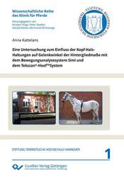
Eine Untersuchung zum Einfluss der Kopf-Hals-Haltungen auf Gelenkwinkel der Hintergliedmaße mit dem Bewegungsanalysesystem Simi und dem Tekscan®-HoofTMSystem (Band 1)
Anna Kattelans (Autor)Vorschau
Inhaltsverzeichnis, Datei (36 KB)
Leseprobe, Datei (160 KB)
Kurzbeschreibung
Die potentiellen Auswirkungen verschiedener Reitweisen sind nicht nur unter
historischer Betrachtung, sondern insbesondere vor der aktuellen Diskussion der sogenannten Rollkur bzw. Hyperflexion bedeutsam. Deshalb sollte in der vorliegenden Arbeit geklärt werden, welchen Einfluss unterschiedliche Kopf-Hals-Haltungen auf die Gliedmaßenmotorik des Pferdes haben. Die Grundlage dafür lag in einer Kombination kinetischer und kinematischer Untersuchungen bei zehn lahmfreien Pferden, die im Schritt und im Trab in drei unterschiedlichen Kopf-Hals-Positionen auf dem Laufband bewegt wurden.
Es wurde im Rahmen der vorliegenden Arbeit zunächst eine Methode zur kinetischen und kinematischen Bewegungsanalyse beim Pferd mit Hilfe von zwei vergleichsweise kostengünstigen Analysesystemen der Firma SIMI und dem Tekscan®-HoofTM-System entwickelt. Anhand der mit dem Videoanalysesystem gemessenen Kopf-Hals-Winkel erfolgte die Objektivierung der drei gewählten Kopf-Hals-Positionen. Die drei Kopf-Hals-Positionen unterscheiden sich sowohl im Schritt als auch im Trab signifikant voneinander. Den kleinsten atlantooccipitalen sowie den größten cervicothorakalen Winkel wies die tiefe Kopf-Hals-Position (HNP 4) auf. Dabei nimmt der Grad der Flexion sowohl im atlantooccipitalen Gelenk von der freien Kopf-Hals-Position zur relativ aufgerichteten und im cervicothorakalen Gelenk von der freien Kopf-Hals-Position über die relativ aufgerichtete bis zur tiefen Kopf-Hals-Position signifikant zu. Das bedeutet, dass einerseits kein signifikanter Unterschied der Flexion im atlantooccipitalen Bereich zwischen der Hyperflexion und der relativen Aufrichtung erkennbar war andererseits jedoch bei Betrachtung der Summe beider Winkel die Abweichung von der natürlichen Kopf-Hals-Position in der Rollkurposition am größten ist. Die Veränderung der Kopf-Hals-Haltung beeinflusste schließlich bei der hier angewendeten Methodik den cervicothorakalen Winkel in hohem Maße stärker als den atlantooccipitalen Winkel.
Zur Interaktion zwischen Kopf-Hals-Position und Hintergliedmaßenwinkel konnte gezeigt werden, dass das Fesselgelenk in der tiefen Kopf-Hals-Position (HNP 4) im Vergleich zur freien (HNP 1) und relativ aufgerichteten Kopf-Hals-Position (HNP 2) signifikant stärker gestreckt wird und die Bewegungsspanne des Hüftgelenks im Trab signifikant zunimmt. Gleichzeitig führt die tiefe Kopf-Hals-Position zu tendenziellen Vergrößerungen des Hüftgelenkwinkels und Verkleinerungen von Knie- und Sprunggelenkwinkel im Vergleich zu den anderen Kopf-Hals-Positionen. Zusammenfassend lässt sich damit eine Überbelastung der Hintergliedmaße in der HNP 4 vermuten, die durch die kinetischen Untersuchungen dieser Studie gestützt wird.
Die mit Hilfe des Tekscan®-HoofTM-System ermittelte vertikale Belastung der Vordergliedmaße ergab in der freien und natürlichen Kopf-Hals-Position signifikant stärkere Kraftwerte (vertikale Belastung) als in der aufgerichteten (HNP 2) und in der tiefen Kopf-Hals-Position (HNP 4). Aufgrund der kinetischen und kinematischen Analyse wird somit angenommen, dass die Entlastung der Vordergliedmaße in der tiefen Kopf-Hals-Position (HNP 4) mit der Überstreckung im Fesselgelenk der Hintergliedmaße einhergeht und somit durch eine zunehmende Belastung der Hintergliedmaße in der tiefen, zur Brust hingezogenen Kopf-Hals-Position kompensiert wird.
Mit der vorliegenden Studie ist es gelungen, kinetische und kinematische Messungen an Pferden mit einem weitgehend kostengünstigen System (Firma Simi und Tekscan®) durchzuführen.
Mit Hilfe dieser Analysesysteme liegt ein weiterer Beitrag zur Erforschung der Auswirkungen unterschiedlicher Kopf-Hals-Haltungen auf die Gliedmaßenmotorik beim Pferd vor. Die Ergebnisse der kinetischen und kinematischen Untersuchungen zeigen, dass die Kopf-Hals-Haltungen, die mit einer stärkeren Beugung als in der klassischen Kopf-Hals-Position einhergehen in den Fesselgelenken der Hintergliedmaße eine stärkere Hyperextension auslösen als die übrigen Körperhaltungen. Das könnte nicht nur eine Ursache für Erkrankungen des Fesselgelenkes, sondern auch für die gestiegene Zahl der Erkrankungen des Fesselträgerursprungs der Hintergliedmaße in den letzten Dekaden sein.
Description
The potential effects of different riding styles are important not only in historical observations but especially with regard to the current discussion on Hyperflexion or Rollkur.
The aim of this study was to analyze the influence of different head and neck positions on hindlimb biomechanics. The basic research concept was a combination of kinetic and kinematic recordings in ten healthy horses which had to walk and trot on a treadmill in three different head and neck positions. In a first step kinematic and kinetic methods were developed for equine gate analysis with a relatively inexpensive three-dimensional high frequency motion analysis system of Simi as well as hoof pressure system of Tekscan®. This video analysis system allowed the objectivization of the three head and neck positions which were chosen for this analysis. The results illustrate that the three head and neck positions differ significantly from each other in walk and trot. Hyperflexion (HNP 4) showes the smallest angle in the atlantooccipital joint and the largest angle in the cervicothoracal joint. The degree of flexion thereby increases significantly in the atlantooccipital joint from the free to the relatively straight head and neck position as well as in the cervicothoracal joint from the free via the relatively straight to hyperflexion. This implies that on the one hand there is no significant difference of flexion in the atlantooccipital joint between the hyperflexion and the relatively straight head and neck position but that on closer examination of the sum of both angles on the other hand the deviance between the natural head and neck position and the low position (HNP 4) was the greatest. The change in the natural head and neck position, as applied in our method, eventually influenced the cervicothoracal angle to a higher degree than the atlantooccipital angle.
Regarding the interaction between the head and neck positions and the hindlimb kinematics it could be demonstrated that the fetlock joint was significantly hyperextended in the low position in comparison to the other two above mentioned head and neck positions and that at the same time the range of motion in the hip joint increased significantly during trot. Furthermore, the low head and neck position tends to results in an increase of the hip joint angle and a decrease of the knee and stifle angle in comparison to the other head and neck positions.
In summary an overstressing of the hindlimb in hyperflexion can be assumed which is supported by the kinetic measurements of this study. The vertical ground reaction force of the frontlimb measured by using Tekscan®-HoofTM-System was significantly higher in the free head and neck position than in the relatively straight and low position. Based on the kinetic and kinematic analysis it can be hypothesized that the hindlimbs had to compensate the load removal of the frontlimbs and the hyperextension of the hindlimb fetlock in the low head and neck position (HNP 4).
In conclusion this study analyzed the kinetic and kinematic forces in movements of horses with two relatively inexpensive analysis systems (Simi and Tekscan®). With the help of these analysis systems it was possible to publish a further crucial contribution for the analysis of limb kinematics of horses. The results of the kinetic and kinematic analysis show that those head-neck-positions which come along with a stronger flexion than those in classical head-neck-positions cause a higher hyperextension in the hindlimb fetlock joints as the other postures do.
This could in turn not only be the cause for disorders in the fetlock joint but also for the increasing number of proximal suspensory desmitis in hindlimbs of horses over the last decades.
| ISBN-13 (Printausgabe) | 9783954041138 |
| ISBN-13 (E-Book) | 9783736941137 |
| Buchendformat | A5 |
| Sprache | Deutsch |
| Seitenanzahl | 154 |
| Umschlagkaschierung | matt |
| Auflage | 1. Aufl. |
| Buchreihe | Wissenschaftliche Reihe der Klinik für Pferde |
| Band | 1 |
| Erscheinungsort | Göttingen |
| Promotionsort | Hannover |
| Erscheinungsdatum | 23.05.2012 |
| Allgemeine Einordnung | Dissertation |
| Fachbereiche |
Veterinärmedizin
|


