| Fachbereiche | |
|---|---|
| Buchreihen (97) |
1382
|
| Nachhaltigkeit |
3
|
| Gesundheitswesen |
1
|
| Geisteswissenschaften |
2372
|
| Naturwissenschaften |
5408
|
| Mathematik | 228 |
| Informatik | 320 |
| Physik | 980 |
| Chemie | 1364 |
| Geowissenschaften | 131 |
| Humanmedizin | 243 |
| Zahn-, Mund- und Kieferheilkunde | 10 |
| Veterinärmedizin | 108 |
| Pharmazie | 147 |
| Biologie | 835 |
| Biochemie, Molekularbiologie, Gentechnologie | 121 |
| Biophysik | 25 |
| Ernährungs- und Haushaltswissenschaften | 45 |
| Land- und Agrarwissenschaften | 1005 |
| Forstwissenschaften | 201 |
| Gartenbauwissenschaft | 20 |
| Umweltforschung, Ökologie und Landespflege | 148 |
| Ingenieurwissenschaften |
1798
|
| Allgemein |
98
|
|
Leitlinien Unfallchirurgie
5. Auflage bestellen |
|
Erweiterte Suche
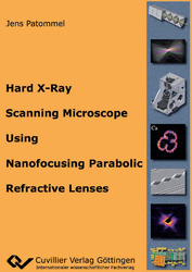
Hard X-Ray Scanning Microscope Using Nanofocusing Parabolic Refractive Lenses
Jens Patommel (Autor)Vorschau
Inhaltsverzeichnis, Datei (47 KB)
Leseprobe, Datei (200 KB)
Hard x rays come along with a variety of extraordinary properties which make them an excellent probe for investigation in science, technology and medicine. Their large attenuation length in matter opens up the possibility to use hard x-rays for non-destructive investigation of the inner structure of specimens. Medical radiography is one important example of exploiting this feature. Since their discovery by W. C. Röntgen in 1895, a large variety of x-ray analytical techniques have been developed and successfully applied, such as x-ray crystallography, reflectometry, fluorescence spectroscopy, x-ray absorption spectroscopy, small angle x-ray scattering, and many more. Each of those methods reveals information about certain physical properties, but usually, these properties are an average over the complete sample region illuminated by the x rays. In order to obtain the spatial distribution of those properties in inhomogeneous samples, scanning microscopy techniques have to be applied, screening the sample with a small x-ray beam. The spatial resolution is limited by the finite size of the beam. The availability of highly brilliant x-ray sources at third generation synchrotron radiation facilities together with the development of enhanced focusing x-ray optics made it possible to generate increasingly small high intense x-ray beams, pushing the spatial resolution down to the sub-100nm range. During this thesis the prototype of a hard x-ray scanning microscope utilizing microstructured nanofocusing lenses was designed, built, and successfully tested. The nanofocusing x-ray lenses were developed by our research group of the Institute of Structural Physics at the Technische Universität Dresden. The prototype instrument was installed at the ESRF beamline ID 13. A wide range of experiments like fluorescence element mapping, fluorescence tomography, x-ray nano-diffraction, coherent x-ray diffraction imaging, and x-ray ptychography were performed as part of this thesis. The hard x-ray scanning microscope provides a stable x-ray beam with a full width at half maximum size of 50–100nm near the focal plane. The nanoprobe was also used for characterization of nanofocusing lenses, crucial to further improve them. Based on the experiences with the prototype, an advanced version of a hard x-ray scanning microscope is under development and will be installed at the PETRA III beamline P06 dedicated as a user instrument for scanning microscopy. This document is organized as follows. A short introduction motivating the necessity for building a hard x-ray scanning microscope is followed by a brief review of the fundamentals of hard x-ray physics with an emphasis on free-space propagation and interaction with matter. After a discussion of the requirements on the x-ray source for the nanoprobe, the main features of synchrotron radiation from an undulator source are shown. The properties of the nanobeam generated by refractive x-ray lenses are treated as well as a two-stage focusing scheme for tailoring size, flux and the lateral coherence properties of the x-ray focus. The design and realization of the microscope setup is addressed, and a selection of experiments performed with the prototype version is presented, before this thesis is finished with a conclusion and an outlook on prospective plans for an improved microscope setup to be installed at PETRA III.
| ISBN-13 (Printausgabe) | 3869556145 |
| ISBN-13 (Printausgabe) | 9783869556147 |
| ISBN-13 (E-Book) | 9783736936140 |
| Buchendformat | A5 |
| Sprache | Englisch |
| Seitenanzahl | 164 |
| Umschlagkaschierung | glänzend |
| Auflage | 1 Aufl. |
| Band | 0 |
| Erscheinungsort | Göttingen |
| Promotionsort | TU Dresden |
| Erscheinungsdatum | 24.01.2011 |
| Allgemeine Einordnung | Dissertation |
| Fachbereiche |
Physik
|


