
Cuvillier Verlag GmbH



| Departments | |
|---|---|
| Book Series (99) |
1415
|
| Nachhaltigkeit |
3
|
| Gesundheitswesen |
3
|
| Humanities |
2412
|
| Natural Sciences |
5430
|
| Mathematics | 230 |
| Informatics | 320 |
| Physics | 982 |
| Chemistry | 1371 |
| Geosciences | 131 |
| Human medicine | 246 |
| Stomatology | 10 |
| Veterinary medicine | 112 |
| Pharmacy | 147 |
| Biology | 837 |
| Biochemistry, molecular biology, gene technology | 121 |
| Biophysics | 25 |
| Domestic and nutritional science | 45 |
| Agricultural science | 1006 |
| Forest science | 201 |
| Horticultural science | 20 |
| Environmental research, ecology and landscape conservation | 149 |
| Engineering |
1821
|
| Common |
97
|
|
Leitlinien Unfallchirurgie
5. Auflage bestellen |
|
Advanced Search
Zeitschätzung, Gedächtnis und Schizophrenie (English shop)
Igor Nenadic (Author)Preview
Table of Contents, Datei (33 KB)
Extract, Datei (59 KB)
Schizophrenia is a complex neuropsychiatric disorder associated with various psychopathological phenomena, in particular disturbances of thought, perception and affectivity. This also includes a pattern of cognitive deficits comprising disturbances of attention and working memory. Numerous studies postulate damage and resulting dysfunction within a network of several cortical and subcortical structures, in particular the hippocampus, thalamus and prefrontal cortex. The dysfunction of prefrontal areas has become the focus of several studies using imaging techniques; however, the finding of “hypofrontality” has not been able to satisfactorily clarify the anatomical focus of prefrontal pathology and its relation to cognitive deficits.
The present investigation takes as its starting point the disturbance of temporal structure in perception and memory in schizophrenia, in particular the impaired ability to process temporal information such as duration or order.
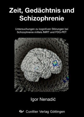
In the first study, functional magnetic resonance imaging was used to depict a neuronal network mediating the ability to estimate time in the range of approximately one second. It could first be shown in healthy subjects that activations in dorsolateral prefrontal, anterior cingulate and superior temporal cortices, as well as in the thalamus and caudate nucleus, occur during time estimation in a manner similar to that in a comparable working memory task (frequency estimation). A region in the right putamen, however, showed time-specific activity and thus represents a potential neuronal correlate of central temporal processing.
Compared with healthy control subjects, schizophrenic patients show reduced activation of the prefrontostriatal network in both tasks. Time-specific hypoactivations are found both in the putamen and in the medial prefrontal cortex and thalamus, which is interpreted as a combined disturbance of basal temporal processing mechanisms in the striatum as well as of prefrontostriatal working memory networks.
In a further study, positron emission tomography was used to examine cortical glucose metabolism during a verbal-serial memory task in patients with schizophrenia or schizotypal personality disorder, as well as in healthy control subjects. An atlas-based procedure for data evaluation allowed the analysis of individual cortical Brodmann areas and their activity patterns. Patients with schizophrenia show a deficit in the recall of semantic information and an increased reliance on serial cognitive strategies as compensation. At the neuronal level this is accompanied by hypometabolism in several prefrontal areas, in particular the dorsolateral, ventrolateral and medial prefrontal cortex, but not the orbitofrontal cortex; for the frontopolar cortex (Brodmann area 10) schizophrenic patients even show higher activation. The pattern of disturbed glucose metabolism can be explained by a disturbance in prefrontal and temporal regions. Drawing on a model by Goldman-Rakic, prefrontal regions for the processing of verbal information also appear to be disproportionately more strongly affected than those for the processing of spatial information. The pattern of changes cannot be explained by a differential reduction of metabolism across different types of cortical areas, which contradicts the postulate of a primary impairment of heteromodal association cortices in schizophrenia.
Patients with schizotypal personality disorder show less pronounced reductions of cortical glucose metabolism, the pattern of which largely follows that of the schizophrenic patients. Deviations are found for medial temporal areas and Brodmann area 10 (frontopolar cortex), for which these patients show increased activity.
Overall, the results demonstrate multifocal frontal functional disturbances in schizophrenia and – less pronounced – in schizotypal personality disorder, which may be regarded as the neuronal basis of disturbed temporal structure in memory. The pattern of cortical functional deficits further appears to underlie the differing degrees of severity of the disturbances within the “schizophrenic spectrum”.
| ISBN-13 (Printausgabe) | 3867274894 |
| ISBN-13 (Hard Copy) | 9783867274890 |
| ISBN-13 (eBook) | 9783736924895 |
| Final Book Format | B5 |
| Language | German |
| Page Number | 114 |
| Edition | 1 |
| Volume | 0 |
| Publication Place | Göttingen |
| Place of Dissertation | Jena |
| Publication Date | 2008-01-17 |
| General Categorization | Dissertation |
| Departments |
Human medicine
|
| Keywords | Schizophrenia, psychology. |


