
Cuvillier Verlag GmbH



| Departments | |
|---|---|
| Book Series (99) |
1415
|
| Nachhaltigkeit |
3
|
| Gesundheitswesen |
3
|
| Humanities |
2411
|
| Natural Sciences |
5429
|
| Mathematics | 230 |
| Informatics | 320 |
| Physics | 982 |
| Chemistry | 1371 |
| Geosciences | 131 |
| Human medicine | 246 |
| Stomatology | 10 |
| Veterinary medicine | 112 |
| Pharmacy | 147 |
| Biology | 837 |
| Biochemistry, molecular biology, gene technology | 121 |
| Biophysics | 25 |
| Domestic and nutritional science | 45 |
| Agricultural science | 1005 |
| Forest science | 201 |
| Horticultural science | 20 |
| Environmental research, ecology and landscape conservation | 148 |
| Engineering |
1821
|
| Common |
97
|
|
Leitlinien Unfallchirurgie
5. Auflage bestellen |
|
Advanced Search
Das Basalapparat-Subproteom von Chlamydomonas reinhardtii (English shop)
Kerstin Brachhold (Author)Preview
Table of Contents, Datei (49 KB)
Extract, Datei (69 KB)
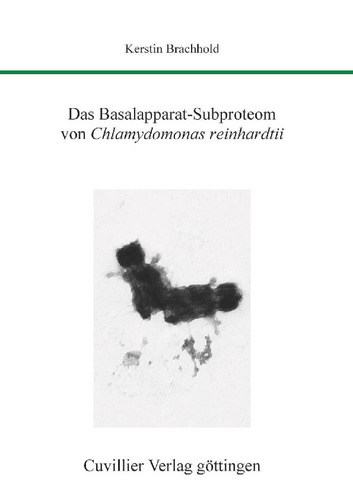
The basal apparatus of the unicellular green alga Chlamydomonas reinhardtii consists of the two flagella-bearing basal bodies as well as two probasal bodies and associated fibres and microtubules. The ultrastructural organisation of the basal bodies largely corresponds to that of the centrioles of animal centrosomes: nine microtubule triplets arranged in a ring form a cylinder approximately 400 nm in length. In cross-sections, various structures can be distinguished, such as the “cartwheel” in the proximal region of the basal bodies or the stellate structure in the transition region to the flagellum. The two basal bodies are linked to each other by connecting fibres and are attached to the plasma membrane by the “transitional fibers”. Starting from the basal bodies, the so-called flagellar roots extend into the cell interior. They connect the basal apparatus with various cell organelles and are thus involved in the internal organisation of the entire cell. As little was previously known about the protein composition of the basal apparatus, the aim of this work was to characterise the proteome of the basal apparatus of C. reinhardtii in more detail.
To identify the proteins, a purification method for basal apparatuses of C. reinhardtii was first developed. The purified basal apparatuses were then separated electrophoretically by 1D SDS-PAGE, the complete gel was cut into 51 bands and the bands were analysed by mass spectrometry. This first approach yielded a total of 1123 different peptides. The peptides could be assigned to 124 gene models, but also to EST entries as well as to genomic positions at which no gene models had previously been located. In a second approach, a basal apparatus pellet was analysed directly – without prior electrophoretic separation – using the MudPIT procedure (“multidimensional protein identification technology”). In this approach, 450 peptides in 295 gene models were identified. In addition, basal apparatuses were reproducibly separated into approximately 60–75 spots by means of 2D-PAGE.
Since a publication on the proteome of the basal bodies of C. reinhardtii was already available at that time (likewise determined by MudPIT), a direct comparison of the respective identified gene models was possible. The comparison of the 295 gene models identified with the MudPIT procedure with this publication yielded a list of 35 partly known (such as α- and β-tubulin, centrin) and new, potential basal apparatus proteins.
Subsequently, five of these new, potential basal apparatus proteins, which had been identified with several peptides in the different approaches (1D gel approach and MudPIT procedure), were characterised in more detail. One of these candidates is erroneously annotated as a mitochondrial translation factor; here, two different genes were presumably combined into one model. For this candidate, a genomic sequence gap was closed. For the four other candidates, partial cDNAs were cloned and initially sequenced. The comparison of these sequences with the gene models led, in the case of candidate 90, to the discovery of two new exons. In the case of candidate 27, it turned out that exon 6 is 42 bp longer than indicated in the gene model. The cDNAs were overexpressed in E. coli and the recombinant proteins were used to generate polyclonal antibodies in rabbits. In Western blots, the antibodies reacted both with the recombinant protein from the bacteria and with the corresponding proteins in isolated basal apparatuses. Using the antibodies, three of the proteins could be localised to the region of the basal apparatus by immunofluorescence microscopy.
| ISBN-13 (Printausgabe) | 3867271887 |
| ISBN-13 (Hard Copy) | 9783867271882 |
| ISBN-13 (eBook) | 9783736921887 |
| Final Book Format | A5 |
| Language | German |
| Page Number | 162 |
| Edition | 1 |
| Volume | 0 |
| Publication Place | Göttingen |
| Place of Dissertation | Köln |
| Publication Date | 2007-03-15 |
| General Categorization | Dissertation |
| Departments |
Biology
|
| Keywords | Basal apparatus, basal bodies, centrioles, proteome analysis, Chlamydomonas reinhardtii. |


