| Departments | |
|---|---|
| Book Series (96) |
1379
|
| Nachhaltigkeit |
3
|
| Gesundheitswesen |
1
|
| Humanities |
2367
|
| Natural Sciences |
5407
|
| Mathematics | 229 |
| Informatics | 319 |
| Physics | 980 |
| Chemistry | 1364 |
| Geosciences | 131 |
| Human medicine | 243 |
| Stomatology | 10 |
| Veterinary medicine | 108 |
| Pharmacy | 147 |
| Biology | 835 |
| Biochemistry, molecular biology, gene technology | 121 |
| Biophysics | 25 |
| Domestic and nutritional science | 45 |
| Agricultural science | 1004 |
| Forest science | 201 |
| Horticultural science | 20 |
| Environmental research, ecology and landscape conservation | 148 |
| Engineering |
1793
|
| Common |
98
|
|
Leitlinien Unfallchirurgie
5. Auflage bestellen |
|
Advanced Search
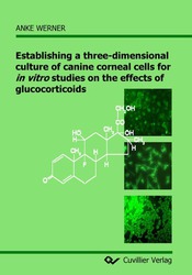
Establishing a three-dimensional culture of canine corneal cells for in vitro studies on the effects of glucocorticoids (English shop)
Anke Werner (Author)Preview
Table of Contents, Datei (21 KB)
Extract, Datei (780 KB)
To provide a model to be used for in vitro studies on drug effects in dogs, the aim of this study was the establishing of a protocol for the primary culture of canine corneal cells (i.e. endothelium, keratocytes, and epithelium) and subsequently the construction of a three¬dimensional culture of canine corneal cells (cornea equivalent). To study the glucocorticoid effects on the three major cell types of the cornea, dexamethasone was used. Since difficulties in the culture of primary canine corneal cells arose, a rabbit epithelial cell line (RCE cell) was used additionally. Both cell types were compared in their reaction to LPS and SDS stimulation, effects of dexamethasone and morphological appearance on the cornea equivalent.
Canine corneal cells were isolated using a combined enzymatic and mechanical technique. In culture, the different cell types were verified with phase contrast microscopy, immunofluorescense and western blotting. The cornea equivalent was constructed step by step in membrane inserts of a six-well plate. Stromal fibroblast in a collagen matrix were seeded onto a confluent endothelial cell layer and cultured for 6 – 8 days. Then either primary canine epithelial cells or RCE cells were added and grown to confluence in a submerged culture. Finally, the equivalent was lifted to the air-liquid-interface for two more weeks to allow a differentiation of the epithelial cells. The glucocorticoid receptor (GR) was investigated in the primary canine corneal cells and the equivalents using RT-PCR and immunohistochemistry. The single cells (including RCE cells) and the cornea equivalents were stimulated with lipopolysaccharide (LPS) and sodium dodecyl sulfate (SDS) and treated with dexamethasone (1.0 and 0.01 µg/ml). The PGE2 concentration, which increases during an inflammatory reactions, was chosen as an indicator to study the effects of dexamethasone after such stimulation. This pro-inflammatory molecule was studied in the culture medium of single cultures of the three major cell types of the canine cornea, of RCE cells as well as in the canine cornea equivalents.
A protocol for the isolation and culture of canine corneal cells was successfully established in this study, and the identity of the cells was verified. The three cell types were successfully reassembled in a vital cornea equivalent which was cultured for a total of five weeks. The GR was detected in both the cultured canine cells and the canine cornea equivalents. The use of RCE cells instead of canine corneal cells in the construction of the cornea equivalents revealed morphological differences. The equivalents constructed with primary canine cells resembled the canine cornea in vivo more closely. An increased PGE2 concentration was measured in canine epithelial cells and keratocytes after the stimulation with LPS and in canine epithelial cells and endothelial following stimulation with SDS. Dexamethasone reduced the LPS-induced PGE2 production in a dose¬dependent manner. The SDS-induced PGE2 concentration was less clearly reduced by dexamethasone, caused by a higher variance of the results. The RCE cells did not react similarly to the primary canine epithelial cells since both stimuli failed to induce an increase in PGE2. In the cornea equivalents, both stimuli led to a significant increase in PGE2 which could be reduced in a dose-dependent manner by both dexamethasone concentrations tested.
The primary culture of the canine corneal cells and the cornea equivalent are interesting systems to test drug effects on corneal cells. The cornea equivalents were even more sensitive to the stimulation and dexamethasone treatment than the single cell cultures. Studies using single cell cultures and the equivalent may reveal further insights on pathophysiological and therapeutic mechanisms in ocular disease. As the dog is one of the species most often treated in veterinary ophthalmology, such models should help improve the treatment of ocular disorders in this species.
| ISBN-13 (Printausgabe) | 3867274517 |
| ISBN-13 (Hard Copy) | 9783867274517 |
| ISBN-13 (eBook) | 9783736924512 |
| Final Book Format | A5 |
| Language | English |
| Page Number | 178 |
| Edition | 1 |
| Volume | 0 |
| Publication Place | Göttingen |
| Place of Dissertation | Hannover |
| Publication Date | 2007-12-11 |
| General Categorization | Dissertation |
| Departments |
Human medicine
|
| Keywords | canin corneal, glucocorticoids, in vito. |


